
Bassett Collection of Stereoscopic Images of Human Anatomy
Creative Commons
Stanford holds the copyright to the David L. Bassett anatomical images and has assigned Creative Commons license Attribution-Share Alike 4.0 International to all of the images.

For additional information regarding use and permissions, please contact the Medical History Center.
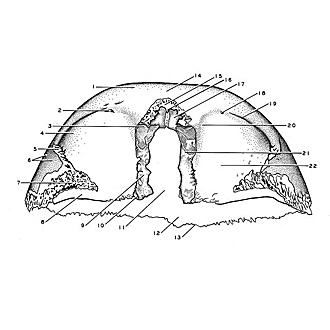
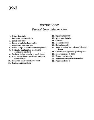
Osteology
Frontal bone, inferior view
- Frontal tuber
- Supraorbital foramen
- Frontal sinus
- Fossa of lacrimal gland
- Zygomatic process
- Temporal line & temporal surface
- Articular surface for greater wing sphenoid bone
- Surface facing middle cranial fossa
- Area which forms roof over ethmoidal cells
- Posterior ethmoidal foramen
- Ethmoidal incisure
- Frontal bone (squamous part)
- Parietal margin
- Glabella
- Nasal margin
- Frontal spine
- Area forming part of roof of nasal fossa
- Canal opening into diploic space
- Supraorbital margin
- Trochlear pit
- Anterior-ethmoidal foramen
- Orbital surface