Bassett Collection of Stereoscopic Images of Human Anatomy
Osteology
Interior of base of skull; middle cranial fossa
Image #36-7
KEYWORDS: Bones cartilage joints.
Creative Commons
Stanford holds the copyright to the David L. Bassett anatomical images and has assigned Creative Commons license Attribution-Share Alike 4.0 International to all of the images.

For additional information regarding use and permissions, please contact the Medical History Center.

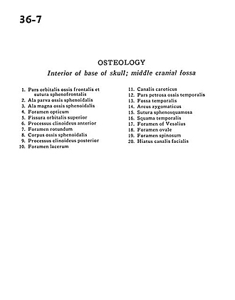
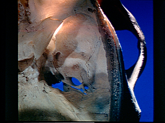
Osteology
Interior of base of skull; middle cranial fossa
- Orbital part of frontal bone &sphenofrontal suture
- Lesser wing sphenoid bone
- Greater wing of sphenoid
- Optic foramen
- Superior orbital fissure
- Anterior clinoid process
- Foramen rotundum
- Sphenoid body bone
- Posterior clinoid process
- Foramen lacerum
- Carotid canal
- Petrosal part of temporal bone
- Temporal fossa
- Zygomatic arch
- Sphenosquamous suture
- Temporal bone (squamous part)
- Foramen of Vesalius
- Foramen ovale
- Foramen spinosum
- Hiatus facial canal