Bassett Collection of Stereoscopic Images of Human Anatomy
Creative Commons
Stanford holds the copyright to the David L. Bassett anatomical images and has assigned Creative Commons license Attribution-Share Alike 4.0 International to all of the images.

For additional information regarding use and permissions, please contact the Medical History Center.


Osteology
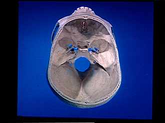
Interior of base of skull
- Crista gall & lamina cribrosa ethmoid bone
- Orbital part of frontal bone & sphenofrontal suture
- Lesser wing sphenoid bone
- Greater wing sphenoid bone
- Sphenosquamous suture
- Temporal bone (squamous part)
- Carotid canal & foramen lacerum
- Petrosquamous fissure
- Petrosal part of temporal bone
- Squamous suture
- Occipitomastoid suture
- Mastoid part of temporal bone
- Occipital bone (squamous part)
- Parietal bone
- Lambdoidal suture
- Foramen magnum
- Internal occipital crest (cruciate eminence)
- Anterior cranial fossa
- Optic foramen & anterior clinoid process
- Sella turcica & posterior clinoid process
- Foramen ovale
- Foramen spinosum
- Middle cranial fossa
- Clivus
- Condyloid canal
- Posterior cranial fossa
- Intersutural bone