Bassett Collection of Stereoscopic Images of Human Anatomy
Osteology
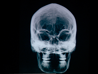
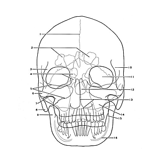
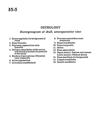
Roentgenogram of skull, anteroposterior view
Image #35-5
KEYWORDS: Bones cartilage joints, Cheek, Eye, Face, Mouth, Nose, Overview.
Creative Commons
Stanford holds the copyright to the David L. Bassett anatomical images and has assigned Creative Commons license Attribution-Share Alike 4.0 International to all of the images.

For additional information regarding use and permissions, please contact the Medical History Center.
Osteology
Roentgenogram of skull, anteroposterior view
- Sagittal suture (in background of view)
- Frontal sinus
- Zygomatic process of frontal bone
- Ethmoidal cells (sella turcica and clinoid processes lie posterior to this area)
- Borders of petrosal part of temporal bone
- Zygomatic arch
- Mandibular articulation
- Mastoid process
- Ramus of mandible
- Temporal fossa
- Orbit
- Sphenoid sinus
- Upper pointer: Bony nasal septum Lower pointer: Hard palate
- Maxillary sinus (in foreground)
- Lingula of mandible
- Mandibular canal