Bassett Collection of Stereoscopic Images of Human Anatomy
Radiographs of the brain
Left internal carotid angiogram, venous phase
Image #34-2
KEYWORDS: Bones cartilage joints, Vasculature, Overview.
Creative Commons
Stanford holds the copyright to the David L. Bassett anatomical images and has assigned Creative Commons license Attribution-Share Alike 4.0 International to all of the images.

For additional information regarding use and permissions, please contact the Medical History Center.
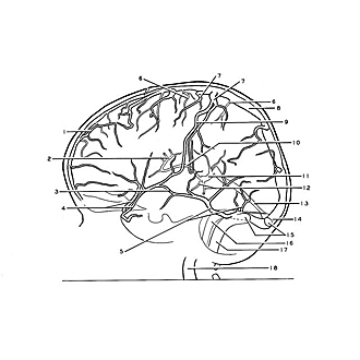
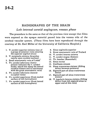
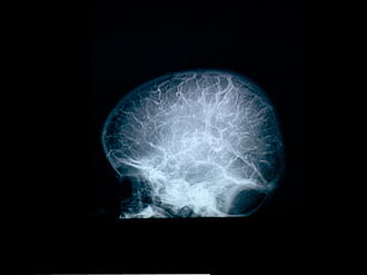
Radiographs of the brain
Left internal carotid angiogram, venous phase
The procedure is the same as that of the previous view except that films were exposed as the opaque material passed into the venous side of the cerebral vascular system. (These films have been reproduced through the courtesy of Dr. Earl Miller of the University of California Hospital.)
- Superior cerebral vein left (one of a group of anterior veins entering the superior sagittal sinus)
- Terminal vein left (branches ramify upon caudate nucleus)
- Small anastomotic vein of Labbé
- Inferior cerebral vein (lower pointer indicates the middle cerebral vein, which lies along the lateral cerebral fissure and is continuous with the great anastomotic vein)
- Inferior cerebral vein (lateral occipital veins)
- Superior cerebral veins (from medial surface of left hemisphere)
- Superior cerebral veins (from lateral surface of left hemisphere)
- Superior sagittal sinus
- Great anastomotic vein of Trolard
- Internal cerebral vein left
- Great cerebral vein of Galen
- Basal veins
- Straight sinus
- Confluence of the sinuses
- Transverse sinus left (filling defect between pointers)
- Sigmoid part of transverse sinus right
- Sigmoid part of transverse sinus left
- Internal jugular vein left (filling defect from left sigmoid sinus to upper end of this vein)