Bassett Collection of Stereoscopic Images of Human Anatomy
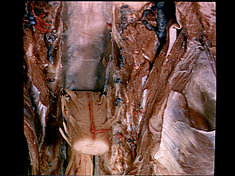
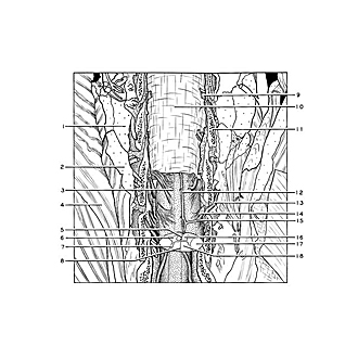
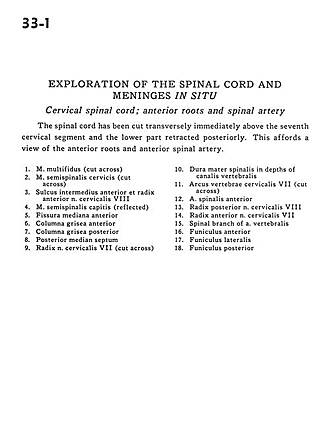
Exploration of the spinal cord and meninges in situ
Cervical spinal cord; anterior roots and spinal artery
Image #33-1
KEYWORDS: Central nervous system, Peripheral nervous system, Vasculature.
Creative Commons
Stanford holds the copyright to the David L. Bassett anatomical images and has assigned Creative Commons license Attribution-Share Alike 4.0 International to all of the images.

For additional information regarding use and permissions, please contact the Medical History Center.
Exploration of the spinal cord and meninges in situ
Cervical spinal cord; anterior roots and spinal artery
The spinal cord has been cut transversely immediately above the seventh cervical segment and the lower part retracted posteriorly. This affords a view of the anterior roots and anterior spinal artery.
- Multifidus muscle (cut across)
- Semispinalis cervicis muscle (cut across)
- Anterior intermediate sulcus and ventral root cervical nerve VIII
- Semispinalis capitis muscle (reflected)
- Ventral median fissure
- Anterior column (gray matter)
- Posterior column (gray matter)
- Posterior median septum
- Root cervical nerve VII (cut across)
- Dura mater in depths of vertebral canal
- Arch of cervical vertebra VII (cut across)
- Anterior spinal artery
- Dorsal root cervical nerve VIII
- Ventral root cervical nerve VII
- Spinal branch of vertebral artery
- Anterior funiculus
- Lateral funiculus
- Posterior funiculus