Bassett Collection of Stereoscopic Images of Human Anatomy
Exploration of the spinal cord and meninges in situ
Dura mater in cervical region
Image #32-4
KEYWORDS: Central nervous system, Meninges.
Creative Commons
Stanford holds the copyright to the David L. Bassett anatomical images and has assigned Creative Commons license Attribution-Share Alike 4.0 International to all of the images.

For additional information regarding use and permissions, please contact the Medical History Center.
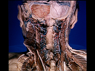
Exploration of the spinal cord and meninges in situ
Dura mater in cervical region
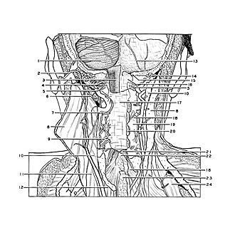
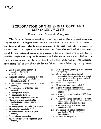
The dura has been exposed by removing part of the occipital bone and the arches of the upper five cervical vertebrae. The cranial dura mater is continuous through the foramen magnum (15) with that which covers the spinal cord. The spinal dura is separated from the wall of the vertebral canal by the epidural space which contains fat and plexiform veins. In the cervical region this space is narrow and the veins are small. Below the foramen magnum the dura is fused with the posterior atlanto-occipital membrane (16) so that above the level of the atlas no epidural space is present.
- Cerebellum (dura removed, arachnoid intact)
- Occipital artery
- Medulla oblongata visible through cerebellomedullary cistern (arachnoid intact)
- Vertebral artery (lying amidst venous plexus)
- Posterior arch of atlas (cut across)
- Greater occipital nerve
- Third occipital nerve
- Splenius capitis muscle (reflected)
- Posterior vertebral venous plexus (between muscle layers)
- Semispinalis capitis muscle (reflected)
- Splenius cervicis muscle (reflected)
- Semispinalis cervicis muscle (cut across)
- Posterior meningeal artery
- Superior obliquus capitis muscle (cut across)
- Occipital bone (at margin of foramen magnum)
- Posterior atlanto-occipital membrane (atlas and the occipital bone to which this was attached have been removed)
- Dura mater (spinal)
- Rotator muscle (cut near origin)
- Posterior vertebral venous plexus (posterior to vertebral arches)
- Cervical vertebral arch IV (cut across)
- Cervical vertebral arch VI with attachment of ligamentum flavum visible along upper margin
- Spinous process cervical vertebra VI
- Spinous process cervical vertebra VII (vertebra prominens)
- Levator scapulae muscle