Bassett Collection of Stereoscopic Images of Human Anatomy
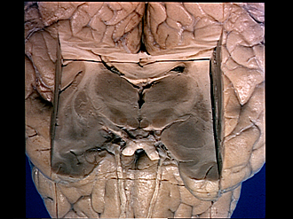
Serial transverse sections of the brain stem
Diencephalon.
Image #31-3
KEYWORDS: Brain, Diencephalon, Midbrain.
Creative Commons
Stanford holds the copyright to the David L. Bassett anatomical images and has assigned Creative Commons license Attribution-Share Alike 4.0 International to all of the images.
For additional information regarding use and permissions, please contact the Medical History Center.
Serial transverse sections of the brain stem
Diencephalon.
A section 4 mm. in thickness has been removed. The tegmental fields (of Forel) are visible at this level.
- Caudate nucleus (tail)
- Circular sulcus
- Third ventricle
- Occipital part internal capsule (posterior limb)
- External medullary lamina (thalamus)
- Thalamic fasciculus (H1 field of Forel)
- Lenticular fasciculus (H2 field of Forel)
- Claustrum
- External capsule
- Lentiform nucleus and posterior part of anterior commissure (lower pointer)
- Hypothalamic nucleus
- Anterior perforated substance and striate arteries
- Optic chiasm
- Corpus callosum
- Lateral ventricle
- Fornix (body)
- Choroid plexus third ventricle and taenia thalami (lower pointer)
- Lateral nucleus of thalamus
- Internal medullary lamina of thalamus
- Medial nucleus of thalamus
- Tegmental field (H field of Forel)
- Mamillary body
- Tuber cinereum
- Amygdaloid nucleus
- Infundibulum (cut across)
- Internal carotid artery


