
Bassett Collection of Stereoscopic Images of Human Anatomy
Serial transverse sections of the brain stem
Pons.
Image #30-6
KEYWORDS: Brain, Cerebellum, Midbrain, Pons, Telencephalon.
Creative Commons
Stanford holds the copyright to the David L. Bassett anatomical images and has assigned Creative Commons license Attribution-Share Alike 4.0 International to all of the images.

For additional information regarding use and permissions, please contact the Medical History Center.

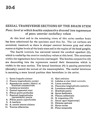
Serial transverse sections of the brain stem
Pons.
At this level and in the remaining views of this series another brain has been substituted for the specimen used thus far. The cut surfaces are unstained, inasmuch as there is sharper contrast between gray and white matter at higher levels of the brain stem and in the region of the basal ganglia.
- Lingual gyrus left
- Longitudinal fissure (cerebral)
- Cerebral aqueduct and brachium conjunctivum (superior cerebellar peduncle)
- Lateral lemniscus
- Central tegmental tract
- Deep pontine fibers
- Longitudinal fasciculus
- Superficial pontine fibers
- Basilar artery
- Internal carotid artery left
- Olfactory tract
- Orbital gyri
- Lobule and ala central lobule
- Locus caeruleus
- Medial longitudinal fasciculus
- Medial lemniscus
- Brachium pontis (middle cerebellar peduncle)
- Fusiform gyrus
- Collateral fissure
- Trigeminal nerve (V)
- Uncus (hippocampal gyrus)
- Optic chiasm
- Straight gyrus
- Temporal pole