
Bassett Collection of Stereoscopic Images of Human Anatomy
Exploration of the meninges and brain in situ
Relation of posterior cerebral artery to tentorium
Image #3-1
KEYWORDS: Brain, Cerebellum, Meninges, Vasculature.
Creative Commons
Stanford holds the copyright to the David L. Bassett anatomical images and has assigned Creative Commons license Attribution-Share Alike 4.0 International to all of the images.

For additional information regarding use and permissions, please contact the Medical History Center.
Exploration of the meninges and brain in situ
Relation of posterior cerebral artery to tentorium
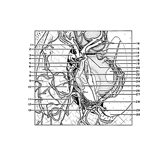
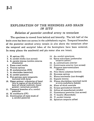
The specimen is viewed from behind and laterally. The left half of the brain stem has been cut across in the subthalamic region. Temporal branches of the posterior cerebral artery remain in situ above the tentorium after the temporal and occipital lobes of the hemisphere have been removed. In many places the arachnoid and pia mater also are intact.
- Optic nerve (II)
- Middle cerebral artery (cut across)
- Internal carotid artery (within chiasmatic cistern)
- Middle cranial fossa
- Optic tract
- Oculomotor nerve (llI)
- Choroidal artery (anterior)
- Posterior cerebral artery
- Petrosal part of temporal bone (covered with dura)
- Upper pointer: Tributaries of basal vein in region of tip of inferior horn of lateral ventricle; Lower pointer: Tentorium cerebelli
- Temporal branches of posterior cerebral artery
- Trochlear nerve (IV)
- Continuation of main trunk of posterior cerebral artery
- Anterior cerebral artery
- Subcallosal gyrus
- Anterior communicating artery
- Anterior commissure (cut across)
- Interventricular foramen (of Monro)
- Free part of column of fornix
- Optic recess
- Massa intermedia (cut through)
- Internal capsule
- Choroid plexus third ventricle and stria medullaris thalami
- Floor of third ventricle
- Lateral geniculate body
- Cerebral auditory aqueduct
- Posterior commissure (cut across)
- Basal vein [Rosenthal]
- Internal cerebral vein
- Corpus callosum (splenium)