Bassett Collection of Stereoscopic Images of Human Anatomy
Serial transverse sections of the brain stem
Medulla oblongata.
Image #29-5
KEYWORDS: Brain, Medulla, Midbrain.
Creative Commons
Stanford holds the copyright to the David L. Bassett anatomical images and has assigned Creative Commons license Attribution-Share Alike 4.0 International to all of the images.

For additional information regarding use and permissions, please contact the Medical History Center.
Serial transverse sections of the brain stem
Medulla oblongata.
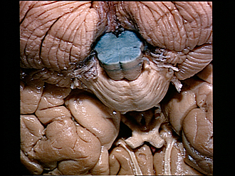
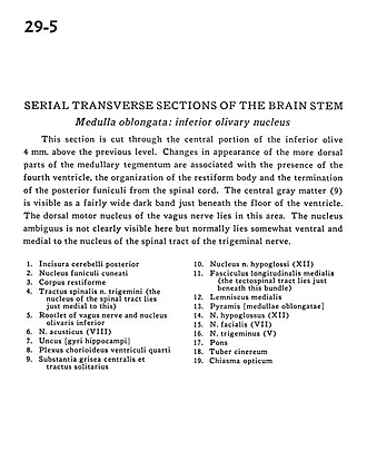
This section is cut through the central portion of the inferior olive 4 mm. above the previous level. Changes in appearance of the more dorsal parts of the medullary tegmentum are associated with the presence of the fourth ventricle, the organization of the restiform body and the termination of the posterior funiculi from the spinal cord. The central gray matter (9) is visible as a fairly wide dark band just beneath the floor of the ventricle. The dorsal motor nucleus of the vagus nerve lies in this area. The nucleus ambiguus is not clearly visible here but normally lies somewhat ventral and medial to the nucleus of the spinal tract of the trigeminal nerve.
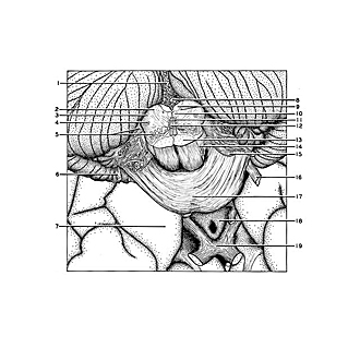
- Posterior cerebellar incisure
- Cuneate nucleus
- Restiform body (inferior cerebellar peduncle)
- Spinal trigeminal tract (the nucleus of the spinal tract lies just medial to this)
- Rootlet of vagus nerve and inferior olivary nucleus
- Vestibulocochlear nerve (VIII)
- Uncus (hippocampal gyrus)
- Choroid plexus fourth ventricle
- Central gray matter and tractus solitarius
- Nucleus hypoglossal nerve (CN XII)
- Medial longitudinal fasciculus (the tectospinal tract lies just beneath this bundle)
- Medial lemniscus
- Pyramid (medulla oblongata)
- Hypoglossal nerve (XII)
- Facial nerve (VII)
- Trigeminal nerve (V)
- Pons
- Tuber cinereum
- Optic chiasm