Bassett Collection of Stereoscopic Images of Human Anatomy
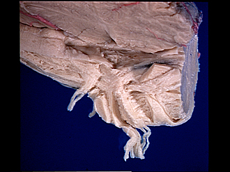
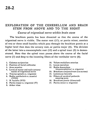
Exploration of the cerebellum and brain stem from above and to the right
Course of trigeminal nerve within brain stem
Image #28-2
KEYWORDS: Brain, Peripheral nervous system, Pons.
Creative Commons
Stanford holds the copyright to the David L. Bassett anatomical images and has assigned Creative Commons license Attribution-Share Alike 4.0 International to all of the images.

For additional information regarding use and permissions, please contact the Medical History Center.
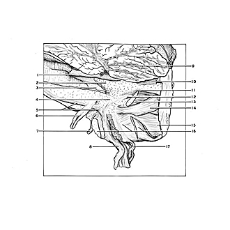
Exploration of the cerebellum and brain stem from above and to the right
Course of trigeminal nerve within brain stem
The brachium pontis has been dissected so that the course of the trigeminal nerve is visible. The motor root (17), or portio minor, consists of two or three small bundles which pass through the brachium pontis at a higher level than does the sensory root, or portio major (8). The division of the latter into a mesencephalic root (12) and a spinal tract (5) is demonstrated. Note that the spinal tract passes above the course of the facial nerve (7) and deep to the incoming fibers of the vestibular nerve (6).
- Calamus scriptorius
- Position of facial colliculus
- Area acustica
- Level of motor and main sensory nuclei of trigeminal nerve
- Spinal trigeminal tract
- Vestibular root of vestibulocochlear nerve (VIII)
- Facial nerve (VII)
- Sensory root trigeminal nerve (V)
- Arbor vitae
- Anterior medullary velum
- Locus caeruleus
- Descending root (mesencephalic) of trigeminal nerve (V)
- Brachium conjunctivum (superior cerebellar peduncle) (cut across)
- Lateral lemniscus
- Fibers of cerebral peduncle entering pons
- Brachium pontis (middle cerebellar peduncle) (dissected)
- Motor root of trigeminal nerve