
Bassett Collection of Stereoscopic Images of Human Anatomy
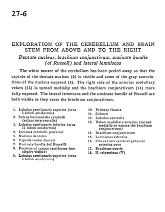
Exploration of the cerebellum and brain stem from above and to the right
Dentate nucleus, brachium conjunctivum, uncinate bundle (of Russell) and lateral lemniscus
Image #27-6
KEYWORDS: Brain, Medulla, Pons.
Creative Commons
Stanford holds the copyright to the David L. Bassett anatomical images and has assigned Creative Commons license Attribution-Share Alike 4.0 International to all of the images.

For additional information regarding use and permissions, please contact the Medical History Center.

Exploration of the cerebellum and brain stem from above and to the right
Dentate nucleus, brachium conjunctivum, uncinate bundle (of Russell) and lateral lemniscus
The white matter of the cerebellum has been peeled away so that the capsule of the dentate nucleus (6) is visible and some of the gray convolutions of the nucleus exposed (5). The right side of the anterior medullary velum (13) is turned medially and the brachium conjunctivum (14) more fully exposed. The lateral lemniscus and the uncinate bundle of Russell are both visible as they cross the brachium conjunctivum.
- Superior semilunar lobe (crus I ansiform lobule)
- Horizontal cerebellar sulcus (sulcus intercruralis)
- Inferior semilunar lobe (crus II ansiform lobule)
- Posterior cerebellar incisure
- Dentate nucleus
- Capsule of dentate nucleus
- Uncinate bundle (of Russell)
- Position of restiform body (inferior cerebellar peduncle) (not clearly visible)
- Superior semilunar lobe (ems I ansiform lobule)
- Primary fissure
- Culmen
- Central lobule
- Anterior medullary velum (turned medially to expose the brachium conjunctivum (superior cerebellar peduncle)
- Brachium conjunctivum (superior cerebellar peduncle)
- Lateral lemniscus
- Fibers from cerebral peduncle entering pons
- Brachium pontis (middle cerebellar peduncle)
- Trigeminal nerve (V)