Bassett Collection of Stereoscopic Images of Human Anatomy
Exploration of the cerebellum and brain stem from the medial aspect
Medullary substance of cerebellar hemisphere
Image #27-4
KEYWORDS: Brain, Cerebellum, Midbrain, Pons.
Creative Commons
Stanford holds the copyright to the David L. Bassett anatomical images and has assigned Creative Commons license Attribution-Share Alike 4.0 International to all of the images.

For additional information regarding use and permissions, please contact the Medical History Center.
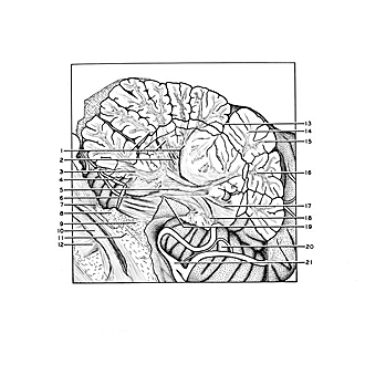
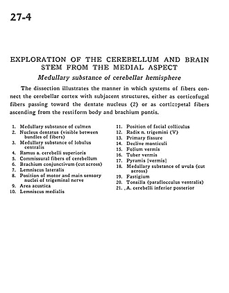
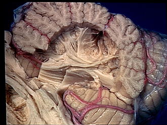
Exploration of the cerebellum and brain stem from the medial aspect
Medullary substance of cerebellar hemisphere
The dissection illustrates the manner in which systems of fibers connect the cerebellar cortex with subjacent structures, either as corticofugal fibers passing toward the dentate nucleus (2) or as corticopetal fibers ascending from the restiform body and brachium pontis.
- Medullary substance of culmen
- Dentate nucleus (visible between bundles of fibers)
- Medullary substance of central lobule
- Branch of superior cerebellar artery
- Commissural fibers of cerebellum
- Brachium conjunctivum (superior cerebellar peduncle) (cut across)
- Lateral lemniscus
- Position of motor and main sensory nuclei of trigeminal nerve
- Area acustica
- Medial lemniscus
- Position of facial colliculus
- Root of trigeminal nerve (V)
- Primary fissure
- Declive of cerebellum
- Folium vermis
- Tuber vermis
- Pyramid (vermis)
- Medullary substance of uvula (cut across)
- Fastigium
- Tonsil (ventral paraflocculus)
- Posterior inferior cerebellar artery