
Bassett Collection of Stereoscopic Images of Human Anatomy
Exploration of the left half of the cerebellum and brain stem from its inferior and medial aspect
Lateral recess of fourth ventricle
Image #25-7
KEYWORDS: Brain, Cerebellum, Ventricules.
Creative Commons
Stanford holds the copyright to the David L. Bassett anatomical images and has assigned Creative Commons license Attribution-Share Alike 4.0 International to all of the images.

For additional information regarding use and permissions, please contact the Medical History Center.

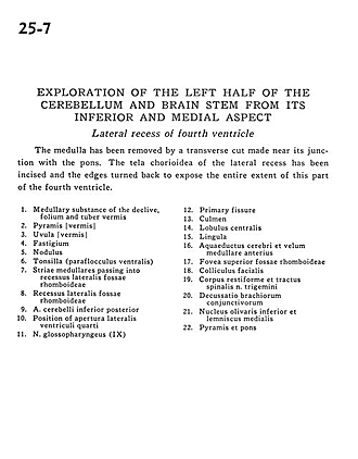
Exploration of the left half of the cerebellum and brain stem from its inferior and medial aspect
Lateral recess of fourth ventricle
The medulla has been removed by a transverse cut made near its junction with the pons. The tela chorioidea of the lateral recess has been incised and the edges turned back to expose the entire extent of this part of the fourth ventricle.
- Medullary substance of the declive,folium and tuber of vermis
- Pyramid (vermis)
- Uvula vermis
- Fastigium
- Nodulus
- Tonsil (ventral paraflocculus)
- Striae medullares passing into lateral recess of rhomboid fossa
- Lateral recess of rhomboid fossa
- Posterior inferior cerebellar artery
- Position of lateral aperture fourth ventricle
- Glossopharyngeal nerve (IX)
- Primary fissure
- Culmen
- Central lobule
- Lingula
- Cerebral aqueduct and anterior medullary velum
- Superior fovea rhomboid fossa
- Facial colliculus
- Restiform body (inferior cerebellar peduncle) and spinal trigeminal tract
- Decussation of superior cerebellar peduncle
- Inferior olivary nucleus and medial lemniscus
- Pyramid and pons