Bassett Collection of Stereoscopic Images of Human Anatomy
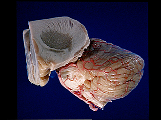
The Surfaces of the cerebellum and brain stem
Lateral aspect
Image #23-4
KEYWORDS: Brain, Cerebellum, Medulla, Pons, Vasculature, Overview.
Creative Commons
Stanford holds the copyright to the David L. Bassett anatomical images and has assigned Creative Commons license Attribution-Share Alike 4.0 International to all of the images.

For additional information regarding use and permissions, please contact the Medical History Center.
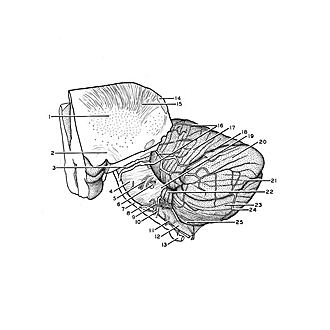
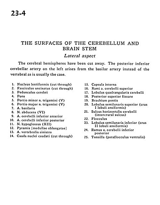
The Surfaces of the cerebellum and brain stem
Lateral aspect
The cerebral hemispheres have been cut away. The posterior inferior cerebellar artery on the left arises from the basilar artery instead of the vertebral as is usually the case.
- Lentiform nucleus (cut through)
- Uncinate fasciculus (cut through)
- Cerebral peduncle
- Pons
- Minor part of trigeminal nerve (V)
- Major part of trigeminal nerve (V)
- Basilar artery
- Abducens nerve (VI)
- Anterior inferior cerebellar artery
- Posterior inferior cerebellar artery
- Hypoglossal nerve (XII)
- Pyramid medulla oblongata
- Vertebral artery left
- Caudate nucleus (tail) (cut through)
- Internal capsule
- Branch of superior cerebellar artery
- Quadrangular lobe of cerebellum
- Posterior superior fissure
- Brachium pontis (middle cerebellar peduncle)
- Superior semilunar lobe (crus I of ansiform lobule)
- Horizontal cerebellar sulcus (intercrural sulcus)
- Flocculus
- Inferior semilunar lobe (crus II ansiform lobule)
- Branch of posterior inferior cerebellar artery
- Tonsil (ventral paraflocculus)