
Bassett Collection of Stereoscopic Images of Human Anatomy
Dissection of anterior aspect of vertebral column
Thoracic region.
Image #220-5
KEYWORDS: Thoracic region, Vertebral column.
Creative Commons
Stanford holds the copyright to the David L. Bassett anatomical images and has assigned Creative Commons license Attribution-Share Alike 4.0 International to all of the images.

For additional information regarding use and permissions, please contact the Medical History Center.
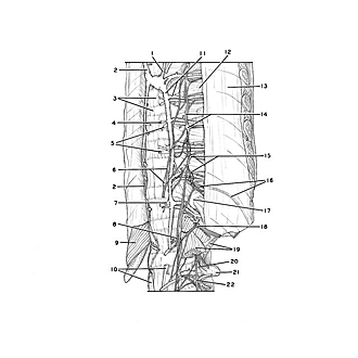

Dissection of anterior aspect of vertebral column
Thoracic region.
The specimen shown in the previous view has been turned to expose its left anterolateral aspect in this close-up view of the lower thoracic and upper lumbar part of the spine.
- Accessory hemiazygos vein
- Azygos vein
- Upper pointer: Body of vertebra Th. VIII (pointer on anterior longitudinal ligament) Lower pointer: Intervertebral disc Th. VII-IX
- Vein draining body of vertebra
- Posterior intercostal arteries IX and X
- Greater splanchnic nerve (lower pointer on splanchnic ganglion)
- Lesser splanchnic nerve
- Least splanchnic it
- Diaphragm (cut off)
- Lumbar part of diaphragm (upper pointer: left crus; lower pointer: right crus)
- Left pointer: Hemiazygos vein Right pointer: Posterior intercostal vein VIII
- Rib VIII
- Costal pleura
- Sympathetic trunk (lower pointer on ganglion of sympathetic trunk)
- Rami communicantes
- Intercostal nerve XI
- Head of rib XII
- Subcostal nerve (note large ramus communicans)
- Upper pointer: Quadratus lumborum muscle Lower pointer: Psoas major muscle
- Ascending lumbar vein
- Transverse process of vertebra L. II
- Lumbar nerve I (passing downward to join lumbar plexus)