
Bassett Collection of Stereoscopic Images of Human Anatomy
Exploration of a brain cut in frontal section at the splenium of the corpus callosum
Roof of third ventricle, habenulae and pineal body
Image #22-1
KEYWORDS: Brain, Diencephalon, Ventricules.
Creative Commons
Stanford holds the copyright to the David L. Bassett anatomical images and has assigned Creative Commons license Attribution-Share Alike 4.0 International to all of the images.

For additional information regarding use and permissions, please contact the Medical History Center.
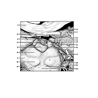

Exploration of a brain cut in frontal section at the splenium of the corpus callosum
Roof of third ventricle, habenulae and pineal body
The internal cerebral veins and their surrounding meninges are now cut away to expose the roof of the third ventricle and the habenulae.
- Corpus callosum
- Hippocampal commissure
- Internal cerebral vein left
- Tela chorioidea third ventricle
- Pineal body
- Superior colliculus
- Brachium of inferior colliculus
- Inferior colliculus
- Trochlear nerve (IV)
- Choroid plexus lateral ventricle
- Fornix (crus) cut across
- Choroidal branch of posterior cerebral artery
- Meninges
- Choroid plexus third ventricle
- Habenula
- Branch of posterior cerebral artery to choroid plexus of third ventricle
- Posterior cerebral artery (cut across)
- Meninges of hippocampal gyrus
- Cerebral peduncle (lateral margin)
- Anterior medullary velum