Bassett Collection of Stereoscopic Images of Human Anatomy
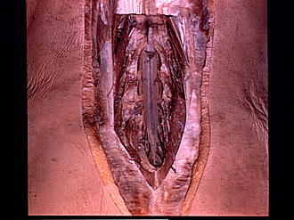
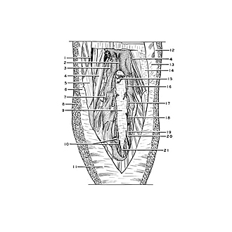
Lumbosacral meninges, spinal cord and nerve roots dissected from behind
Dura mater in lumbar region
Image #219-5
KEYWORDS: Central nervous system, Lumbar region, Sacral region, Vertebral column.
Creative Commons
Stanford holds the copyright to the David L. Bassett anatomical images and has assigned Creative Commons license Attribution-Share Alike 4.0 International to all of the images.

For additional information regarding use and permissions, please contact the Medical History Center.
Lumbosacral meninges, spinal cord and nerve roots dissected from behind
Dura mater in lumbar region
The laminae of the third, fourth and fifth lumbar vertebrae and of the upper two sacral segments have been resected. Epidural fat has been removed to expose segmentally arranges veins (15). Several fibrous strands (10) pass from the lower part of the dura to the sacrum as part of the anchoring mechanism of the meninges.
- Lateral cutaneous branch of dorsal branch thoracic nerve XII
- Spinous process vertebra L. II
- Inferior articular process vertebra L. II
- Longissimus thoracis muscle
- Iliocostalis muscle
- Thoracolumbar fascia
- Multifidus muscle (cut across near origin from mammillary process)
- Superficial fascia
- Dura mater
- Fibrous strands attaching dura to sacrum
- Gluteus maximus muscle
- Fascia of latissimus dorsi muscle
- Aponeurosis of latissimus dorsi muscle
- Layer of fascia beneath thoracolumbar fascia
- Posterior internal vertebral venous plexus
- Laminal arch of vertebra L. III (cut across)
- Laminal arch of vertebra L. IV (cut across)
- Laminal arch of vertebra L. V (cut across)
- Laminal arch of sacrum (cut across)
- Sacral nerve I
- Dural filum terminale (coccygeal nerve roots accompany this band)