Bassett Collection of Stereoscopic Images of Human Anatomy
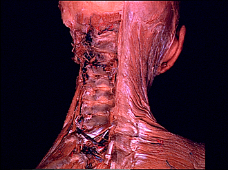
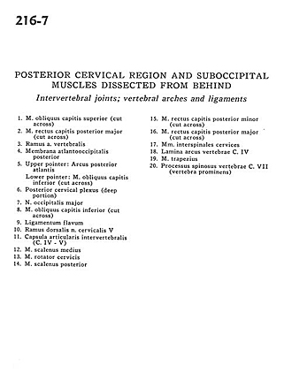
Posterior cervical region and suboccipital muscles dissected from behind
Intervertebral joints; vertebral arches and ligaments
Image #216-7
KEYWORDS: Cervical region, Muscles and tendons, Vertebral column.
Creative Commons
Stanford holds the copyright to the David L. Bassett anatomical images and has assigned Creative Commons license Attribution-Share Alike 4.0 International to all of the images.

For additional information regarding use and permissions, please contact the Medical History Center.
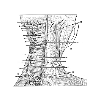
Posterior cervical region and suboccipital muscles dissected from behind
Intervertebral joints; vertebral arches and ligaments
- Superior oblique capitis muscle (cut across)
- Major posterior rectus capitis muscle (cut across)
- Branch of vertebral artery
- Posterior atlanto-occipItal membrane
- Upper pointer: Posterior arch of atlas Lower pointer: Inferior oblique capitis muscle (cut across)
- Posterior cervical plexus (deep portion)
- Greater occipital nerve
- Inferior oblique capitis muscle (cut across)
- Ligamentum flavum
- Dorsal branch cervical nerve V
- Intervertebral joint capsule (C. IV - V)
- Middle scalene muscle
- Rotator cervicis muscle
- Posterior scalene muscle
- Minor posterior rectus capitis muscle (cut across)
- Major posterior rectus capitis muscle (cut across)
- Interspinales cervices muscles
- Lamina (arch of vertebra) C. IV
- Trapezius muscle
- Spinous process vertebra C. VII (vertebra prominens)