Bassett Collection of Stereoscopic Images of Human Anatomy
Posterior cervical region and suboccipital muscles dissected from behind
Suboccipital muscles; suboccipital triangle, left posterolateral view
Image #216-6
KEYWORDS: Cervical region, Muscles and tendons, Vertebral column.
Creative Commons
Stanford holds the copyright to the David L. Bassett anatomical images and has assigned Creative Commons license Attribution-Share Alike 4.0 International to all of the images.

For additional information regarding use and permissions, please contact the Medical History Center.

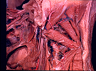
Posterior cervical region and suboccipital muscles dissected from behind
Suboccipital muscles; suboccipital triangle, left posterolateral view
- Superior oblique capitis muscle
- Mastoid cells (dissected)
- Occipital artery
- Longissimus capitis muscle (cut across)
- Muscular branch occipital artery
- Facial nerve (VII)
- Digastric muscle (cut across)
- Posterior cervical plexus (of Cruveilhier)
- Transverse process of atlas
- Inferior oblique capitis muscle
- Hypoglossal nerve (XII)
- Posterior intertransverse muscle
- Intervertebral joint capsule (C. II- III)
- Vagus nerve (X)
- Dorsal branch cervical nerve IV
- Occipital bone
- Minor posterior rectus capitis muscle
- Major posterior rectus capitis muscle
- Nuchal ligament
- Suboccipital nerve (emerging through suboccipital triangle)
- Posterior arch of atlas
- Trapezius muscle
- Greater occipital nerve
- Spinous process axis
- Arch of axis
- Semispinalis cervicis muscle
- Multifidus muscle