Bassett Collection of Stereoscopic Images of Human Anatomy
Dissection of thoracic and lumbosacral regions of back from a posterior approach
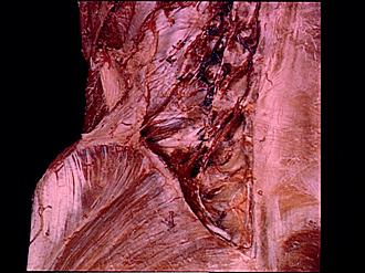
Rotator, interspinal and intertransverse muscles in lumbar region
Image #214-5
KEYWORDS: Lumbar region, Muscles and tendons, Sacral region, Thoracic region, Vertebral column.
Creative Commons
Stanford holds the copyright to the David L. Bassett anatomical images and has assigned Creative Commons license Attribution-Share Alike 4.0 International to all of the images.

For additional information regarding use and permissions, please contact the Medical History Center.


Dissection of thoracic and lumbosacral regions of back from a posterior approach
Rotator, interspinal and intertransverse muscles in lumbar region
The erector spinae and multifidus muscles have been nearly completely excised from the lumbar region.
- Medial intertransverse muscle
- Lateral intertransverse muscle (several fascicles present)
- Dorsal branch thoracic nerve XII
- Rib XII
- Aponeurosis transversus abdominis muscle
- Thoracolumbar fascia (upper pointer, middle layer; lower pointer, posterior layer, cut off)
- Transversus abdominis muscle
- Quadratus lumborum muscle (covered by thoracolumbar fascia)
- Upper pointer: Longissimus muscle Lower pointer: Iliocostalis lumborum muscle
- Iliac crest
- Superior cluneal nerves
- Gluteus medius muscle
- Gluteus maximus muscle
- Posterior superior iliac spine
- Spinous process vertebra L. II
- Intervertebral joint capsule
- Interspinalis lumborum muscle
- Transverse process of vertebra L. IV
- Thoracolumbar fascia (posterior layer)
- Rotator muscles
- Spinous process vertebra L. V
- Upper pointer: Lamina (arch of vertebra) L. V Lower pointer: Intervertebral joint capsule L. V - S. I
- Arch sacrum