Bassett Collection of Stereoscopic Images of Human Anatomy
Dissection of thoracic and lumbosacral regions of back from a posterior approach
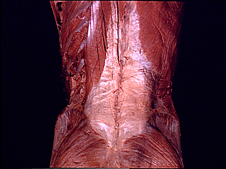
Erector spinae muscle, close-up view of lumbosacral portion
Image #213-4
KEYWORDS: Lumbar region, Muscles and tendons, Sacral region, Thoracic region, Vertebral column.
Creative Commons
Stanford holds the copyright to the David L. Bassett anatomical images and has assigned Creative Commons license Attribution-Share Alike 4.0 International to all of the images.

For additional information regarding use and permissions, please contact the Medical History Center.


Dissection of thoracic and lumbosacral regions of back from a posterior approach
Erector spinae muscle, close-up view of lumbosacral portion
The posterior layer of thoracolumbar fascia (9)has been cut away on the left to reveal the erector spinae muscle.
- Iliocostalis lumborum muscle (insertion)
- Longissimus thoracis muscle
- Spinalis thoracis muscle
- Serratus posterior inferior muscle (cut off near insertion into tenth rib)
- Lateral cutaneous branch of dorsal branch thoracic nerve X
- Rib XII
- Internal oblique muscle (reflected anteriorly)
- Transverse abdominis muscle (aponeurosis of origin)
- Thoracolumbar fascia (cut across)
- Erector spinae muscle
- Superior cluneal nerves
- Latissimus dorsi muscle
- Thoracolumbar fascia (posterior layer)
- External oblique muscle
- Lumbar triangle (Petit's)
- Iliac crest
- Gluteus medius muscle
- Gluteus maximus muscle