Bassett Collection of Stereoscopic Images of Human Anatomy
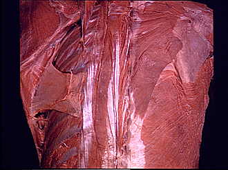
Dissection of thoracic and lumbosacral regions of back from a posterior approach
Erector spinae muscle, close-up view of thoracic portion
Image #213-3
KEYWORDS: Lumbar region, Muscles and tendons, Sacral region, Thoracic region, Vertebral column.
Creative Commons
Stanford holds the copyright to the David L. Bassett anatomical images and has assigned Creative Commons license Attribution-Share Alike 4.0 International to all of the images.

For additional information regarding use and permissions, please contact the Medical History Center.

Dissection of thoracic and lumbosacral regions of back from a posterior approach
Erector spinae muscle, close-up view of thoracic portion
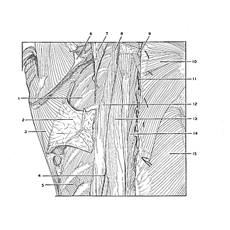
In this close-up view of the upper part of the specimen shown in the preceding photograph of the iliocostalis (12), longissimus (13) and spinalis (14) divisions of the erector spinae are more clearly visible.
- Rib V
- Fascia of serratus anterior muscle
- Serratus anterior muscle
- Lateral cutaneous branch of dorsal branch thoracic nerve IX
- Serratus posterior inferior muscle (insertion into ninth rib)
- Scapula
- Serratus posterior superior muscle (reflected laterally)
- Iliocostalis cervicis muscle
- Spinous process vertebra Th. II
- Trapezius muscle
- Middle cutaneous branch of dorsal branch .thoracic nerve III
- Iliocostalis muscle
- Longissimus thoracis muscle
- Spinalis thoracis muscle
- Latissimus dorsi