Bassett Collection of Stereoscopic Images of Human Anatomy
Dissection of thoracic and lumbosacral regions of back from a posterior approach
Nerve supply to serratus posterior superior muscle
Image #212-7
KEYWORDS: Lumbar region, Muscles and tendons, Sacral region, Thoracic region, Vertebral column.
Creative Commons
Stanford holds the copyright to the David L. Bassett anatomical images and has assigned Creative Commons license Attribution-Share Alike 4.0 International to all of the images.

For additional information regarding use and permissions, please contact the Medical History Center.
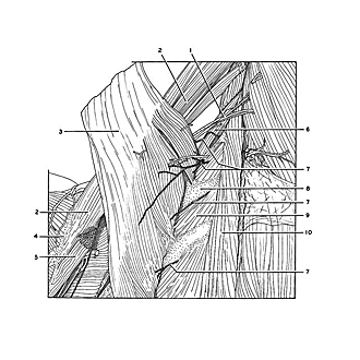
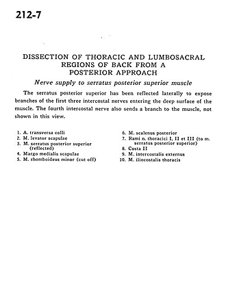
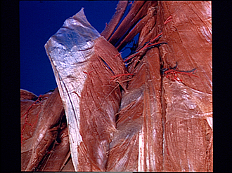
Dissection of thoracic and lumbosacral regions of back from a posterior approach
Nerve supply to serratus posterior superior muscle
The serratus posterior superior has been reflected laterally to expose branches of the first three intercostal nerves entering the deep surface of the muscle. The fourth intercostal nerve also sends a branch to the muscle, not shown in this view.
- Transverse colli artery
- Levator scapulae muscle
- Serratus posterior superior muscle (reflected)
- Medial margin of scapula
- Rhomboid minor muscle (cut off)
- Posterior scalene muscle
- Branches of thoracic nerve I, II and III (to serratus posterior superior muscle)
- Rib II
- External intercostal muscle
- Iliocostalis muscle