
Bassett Collection of Stereoscopic Images of Human Anatomy
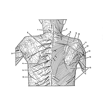
Dissection of thoracic and lumbosacral regions of back from a posterior approach
Superficial structures and external muscles of thoracic region of back
Image #212-2
KEYWORDS: Lumbar region, Muscles and tendons, Peripheral nervous system, Sacral region, Thoracic region, Vasculature, Vertebral column.
Creative Commons
Stanford holds the copyright to the David L. Bassett anatomical images and has assigned Creative Commons license Attribution-Share Alike 4.0 International to all of the images.

For additional information regarding use and permissions, please contact the Medical History Center.
Dissection of thoracic and lumbosacral regions of back from a posterior approach
Superficial structures and external muscles of thoracic region of back
On the lower half of the specimen the tela subcutanea has been dissected to expose cutaneous nerves and vessels. To the right of the midline the trapezius and latissimus dorsi muscles have been exposed.
- Cervical surface of superficial fascia (nuchal fascia)
- Medial dorsal cutaneous branch thoracic nerve III
- Intercostobrachial nerve
- Triangle of auscultation
- Fascia covering latissimus dorsi muscle
- Lateral cutaneous branch intercostal nerve V (posterior branch)
- Spinous process vertebra C. VII
- Trapezius muscle
- Spine of scapula
- Acromion
- Fascia infraspinata
- Deltoid muscle
- Triceps brachii muscle
- Teres major muscle
- Latissimus dorsi muscle