
Bassett Collection of Stereoscopic Images of Human Anatomy
Arteries of vertebral column of one
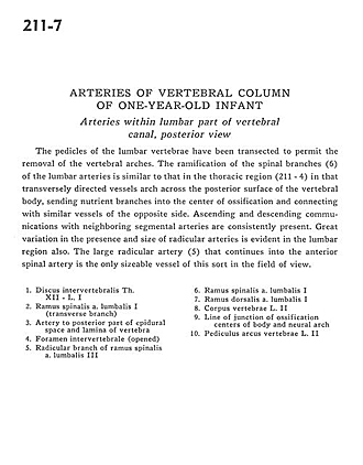
Year-old infant - Arteries of lumbar part of vertibral canal, posterior view
Image #211-7
KEYWORDS: Bones joints cartilage, Cervical region, Thoracic region, Vasculature, Vertebral column.
Creative Commons
Stanford holds the copyright to the David L. Bassett anatomical images and has assigned Creative Commons license Attribution-Share Alike 4.0 International to all of the images.

For additional information regarding use and permissions, please contact the Medical History Center.

Arteries of vertebral column of one
Year-old infant - Arteries of lumbar part of vertibral canal, posterior view
The pedicles of the lumbar vertebrae have been transformed to permit the removal of the vertebral arches. The ramification of the spinal branches (6) of the lumbar arteries is similar to that in the thoracic region (211-4) in that transversely directed vessels arch across the posterior surface of the vertebral body, sending nutrient branches into the center of ossification and connecting with similar vessels of the opposite side. Ascending and descending communications with neighboring segmental arteries are consistently present. Great variation in the presence and size of radicular arteries is evident in the lumbar region also. The large radicular artery (5) that continues into the anterior spinal artery is the only sizeable vessel of this sort in the field of view.
- Intervertebral disc Th. XII - L. I
- Spinal branch lumbar artery I (transverse branch)
- Artery to posterior part of epidural space and lamina of vertebra
- Intervertebral foramen (opened)
- Radicular branch of spinal branch lumbar artery III
- Spinal branch lumbar artery
- Dorsal branch lumbar artery
- Body of vertebra L. II
- Line of junction of ossification centers of body and neural arch
- Pedicle (arch of vertebra) L. II