Bassett Collection of Stereoscopic Images of Human Anatomy
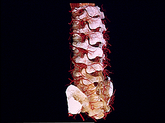
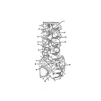
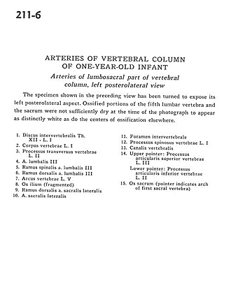
Arteries of vertebral column of one
Year-old infant - Arteries of lumbosacral part of vertebral column, left posterolateral view
Image #211-6
KEYWORDS: Bones joints cartilage, Cervical region, Thoracic region, Vasculature, Vertebral column.
Creative Commons
Stanford holds the copyright to the David L. Bassett anatomical images and has assigned Creative Commons license Attribution-Share Alike 4.0 International to all of the images.

For additional information regarding use and permissions, please contact the Medical History Center.
Arteries of vertebral column of one
Year-old infant - Arteries of lumbosacral part of vertebral column, left posterolateral view
The specimen shown in the preceding view has been turned to expose its left posterolateral aspect. Ossified portions of the fifth lumbar vertebra and the sacrum were not sufficiently dry at the time of the photograph to appear as distinctly white as do the centers of ossification elsewhere.
- Intervertebral disc Th. XII - L. I
- Body of vertebra L. I
- Transverse process of vertebra L. II
- Lumbar artery III
- Spinal branch lumbar artery III
- Dorsal branch lumbar artery III
- Arch of vertebra L. V
- Ilium (fragmented)
- Dorsal branch lateral sacral artery
- Lateral sacral artery
- Intervertebral foramen
- Spinous process vertebra L. 1
- Vertebral canal
- Upper pointer: Superior articular process vertebra L. III Lower pointer: Inferior articular process vertebra L. II
- Sacrum (pointer indicates arch of first sacral vertebra)