Bassett Collection of Stereoscopic Images of Human Anatomy
Radiography
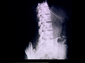
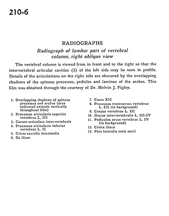
Radiograph of lumbar part of vertebral column, right lateral view
Image #210-6
KEYWORDS: Bones joints cartilage, Lumbar region, Vertebral column.
Creative Commons
Stanford holds the copyright to the David L. Bassett anatomical images and has assigned Creative Commons license Attribution-Share Alike 4.0 International to all of the images.

For additional information regarding use and permissions, please contact the Medical History Center.
Radiography
Radiograph of lumbar part of vertebral column, right lateral view
The vertebral column is viewed from in front and to the right so that the intervertebral articular cavities (3) of the left side may be seen in profile. Details of the articulations on the right side are obscured by the overlapping shadows of hte spinous processes, pedicles and laminae of the arches. This film was obtained through the courtesy of Dr. Melvin J. Figley.
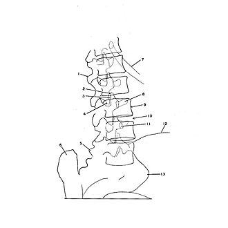
- Overlapping shadows of spinous processes and arches (area indicated extends vertically throughout film)
- Superior articular process vertebra L. III
- Intervertebral articular cavity
- Inferior articular process vertebra L. II
- Intermediate sacral crest
- Ilium
- Rib XII
- Transverse process of vertebra L. III (in background)
- Body of vertebra L. III
- Intervertebral disc L. IIl-IV
- Pedicle (arch of vertebra) L. IV (in background)
- Iliac crest
- Lateral part sacrum