
Bassett Collection of Stereoscopic Images of Human Anatomy
Radiography
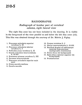
Radiograph of lumbar part of vertebral column, right lateral view
Image #210-5
KEYWORDS: Bones joints cartilage, Lumbar region, Vertebral column.
Creative Commons
Stanford holds the copyright to the David L. Bassett anatomical images and has assigned Creative Commons license Attribution-Share Alike 4.0 International to all of the images.

For additional information regarding use and permissions, please contact the Medical History Center.

Radiography
Radiograph of lumbar part of vertebral column, right lateral view
The right iliac crest has not been included in the drawing. It is visible in the foreground of the view parallel to and below the left iliac crest (13). This film was obtained through the courtesy of Dr. Melvin J. Figley.
- Superior articular process vertebra L. II
- Inferior articular process vertebra L. I
- Pedicle (arch of vertebra) L. II
- Intervertebral articular cavity L. Il-III
- Spinous process vertebra L. III
- Intervertebral foramen
- Superior articular process of sacrum
- Median sacral crest
- Sacral canal
- Body of vertebra L. I
- Intervertebral disc L. II-III
- Residual droplet of radio-opaque material from previous myelographic examination
- Iliac crest (in background)
- Anterior superior iliac spine (in background)
- Promontory