Bassett Collection of Stereoscopic Images of Human Anatomy
Radiography
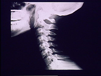
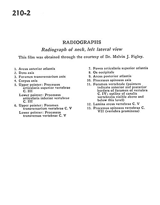
Radiograph of neck, left lateral view
Image #210-2
KEYWORDS: Bones joints cartilage, Cervical region, Vertebral column.
Creative Commons
Stanford holds the copyright to the David L. Bassett anatomical images and has assigned Creative Commons license Attribution-Share Alike 4.0 International to all of the images.

For additional information regarding use and permissions, please contact the Medical History Center.

Radiography
Radiograph of neck, left lateral view
This film was obtained through the courtesy of Dr. Melvin J. Figley.
- Anterior arch of atlas
- Dens (axis)
- Transverse foramen axis
- Body of axis
- Upper pointer: Superior articular process vertebra C. III Lower pointer: Inferior articular process vertebra C. III
- Upper pointer: Transverse foramen vertebra C. V Lower pointer: Transverse process vertebra C. V
- Superior articular facet of atlas
- Occipital bone
- Posterior arch of atlas
- Spinous process axis
- Vertebral foramen (pointers indicate anterior and posterior borders of foramen of vertebra C. IV, outline of vertebral canal visible above and below this level)
- Lamina (arch of vertebra) C. V
- Spinous process vertebra C. VII (vertebra prominens)