Bassett Collection of Stereoscopic Images of Human Anatomy
Exploration of a brain cut in frontal section at the splenium of the corpus callosum
Cingulum, fornix, pulvinar and choroidal arteries
Image #21-6
KEYWORDS: Brain, Diencephalon, Occipital lobe, Telencephalon, Vasculature.
Creative Commons
Stanford holds the copyright to the David L. Bassett anatomical images and has assigned Creative Commons license Attribution-Share Alike 4.0 International to all of the images.

For additional information regarding use and permissions, please contact the Medical History Center.

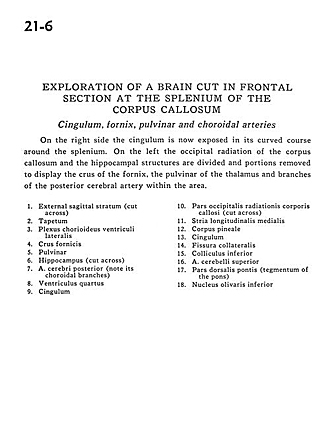
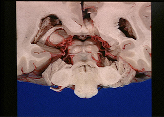
Exploration of a brain cut in frontal section at the splenium of the corpus callosum
Cingulum, fornix, pulvinar and choroidal arteries
On the right side the cingulum is now exposed in its curved course around the splenium. On the left the occipital radiation of the corpus callosum and the hippocampal structures are divided and portions removed to display the crus of the fornix, the pulvinar of the thalamus and branches of the posterior cerebral artery within the area.
- External sagittal stratum (cut across)
- Tapetum
- Choroid plexus lateral ventricle
- Fornix (ems)
- Pulvinar
- Hippocampus (cut across)
- Posterior cerebral artery (note its choroidal branches)
- Fourth ventricle
- Cingulum
- Occipital part of radiations of corpus callosum (cut across)
- Medial longitudinal stria
- Pineal body
- Cingulum
- Collateral fissure
- Inferior colliculus
- Superior cerebellar artery
- Dorsal part of pons (tegmentum of the pons)
- Inferior olivary nucleus