
Bassett Collection of Stereoscopic Images of Human Anatomy
Sagittal sections of right foot
Section through lateral part of foot, viewed from lateral side
Image #205-7
KEYWORDS: Ankle, Foot and toes.
Creative Commons
Stanford holds the copyright to the David L. Bassett anatomical images and has assigned Creative Commons license Attribution-Share Alike 4.0 International to all of the images.

For additional information regarding use and permissions, please contact the Medical History Center.

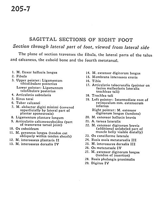
Sagittal sections of right foot
Section through lateral part of foot, viewed from lateral side
The plane of section traverses the fibula, the lateral parts of the talus and calcaneus, the cuboid bone and the fourth metatarsal.
- Flexor hallucis longus muscle
- Fibula
- Upper pointer: Posterior tibiofibular ligament Lower pointer: Posterior talofibular ligament
- Subtalar articulation
- Tarsal sinus
- Tuberosity of calcaneus
- Abductor digiti minimi muscle (covered superficially by lateral part of plantar aponeurosis)
- Long plantar ligament
- Calcaneocuboid articulation (part of transverse tarsal joint)
- Cuboid bone
- Peroneus longus muscle (tendon cut obliquely within tendon sheath)
- 2nd plantar interosseus muscle
- 4th dorsal interosseus muscle
- Extensor digitorum longus muscle
- Interosseous membrane of leg
- Tibia
- Talocrural articulation (pointer on lateral malleolar surface of trochlea of talus)
- Trochlea of talus
- Left pointer: Intermediate root of inferior extensor retinaculum Right pointer: Extensor digitorum longus muscle (tendons)
- Extensor hallucis brevis muscle
- Lateral tarsal artery
- Extensor digitorum brevis muscle (additional unlabeled part of muscle belly visible distally)
- Lateral cuneiform bone
- Base of 3rd metatarsal bone
- 3rd dorsal interosseus muscle
- 4th metatarsal bone
- Extensor digitorum longus muscle (tendon of insertion)
- Base of proximal phalanx
- 4th digit