Bassett Collection of Stereoscopic Images of Human Anatomy
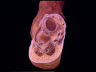
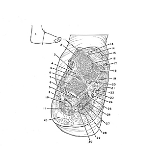
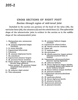
Cross sections of right foot
Section through region of mid-tarsal joint
Image #205-2
KEYWORDS: Bones joints cartilage, Foot and toes.
Creative Commons
Stanford holds the copyright to the David L. Bassett anatomical images and has assigned Creative Commons license Attribution-Share Alike 4.0 International to all of the images.

For additional information regarding use and permissions, please contact the Medical History Center.
Cross sections of right foot
Section through region of mid-tarsal joint
Included in the section are portions of the head of the talus (16), the navicular bone (18), the calcaneus (5) and the cuboid bone (7). The spheroidal shape of the talonavicular joint is evident in the section as is the saddle-shape of the calcaneocuboid joint.
- Inferior extensor retinaculum
- Extensor digitorum longus muscle (tendons)
- Lateral tarsal artery
- Bifurcate ligament
- Calcaneus
- Extensor digitorum brevis muscle
- Cuboid bone
- Long plantar ligament
- Peroneus brevis muscle (tendon)
- Upper pointer: Tendinous sheath of peroneus longus and plantaris muscles Lower pointer: Peroneus longus muscle (sesamoid bone present in tendon)
- Abductor digiti minimi muscle
- Tela subcutanea
- Extensor hallucis longus muscle (tendon)
- Talonavicular ligament
- Tibialis anterior muscle (tendon)
- Head of talus
- Greater saphenous vein
- Navicular bone
- Tibialis posterior muscle (upper pointer indicates sesamoid bone in tendon)
- Plantar calcaneonavicular ligament (spring ligament)
- Abductor hallucis muscle
- Flexor digitorum longus muscle (tendon in common sheath with tendon of flexor hallucis longus)
- Flexor hallucis longus muscle (tendon)
- Medial plantar nerve
- Calcaneocuboid plantar ligament
- Quadratus plantae muscle
- Flexor digitorum brevis muscle
- Lateral plantar nerve
- Plantar aponeurosis
- Lateral plantar artery