Bassett Collection of Stereoscopic Images of Human Anatomy
Cross sections of right foot
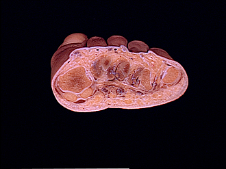
Section of distal part of foot, viewed from behind
Image #204-4
KEYWORDS: Bones joints cartilage, Foot and toes.
Creative Commons
Stanford holds the copyright to the David L. Bassett anatomical images and has assigned Creative Commons license Attribution-Share Alike 4.0 International to all of the images.

For additional information regarding use and permissions, please contact the Medical History Center.


Cross sections of right foot
Section of distal part of foot, viewed from behind
The plane of section passes through the sesamoid bones of the first metatarsophalangeal joint medially and the head of the fifth metatarsal laterally.
- Head of metatarsal bone I (note articular cartilage covering lower part of head)
- Collateral ligament for metatarsophalangeal articulation
- Abductor hallucis muscle (tendon approaching insertion)
- Sesamoid bone
- Flexor hallucis longus muscle (tendon)
- Adductor hallucis muscle (tendon of oblique head)
- Common plantar digital nerve
- Left pointer: 1st lumbrical muscle Right pointer: Flexor digitorum longus and brevis muscles (tendons cut off within fibrous sheath)
- Common plantar digital nerve
- Left pointer: 2nd lumbrical muscle Right pointer: Flexor digitorum longus and brevis muscles (tendons cutoff in sheath)
- Transverse head of adductor hallucis muscle
- Flexor digitorum longus and brevis muscles (to 4th toe)
- Extensor hallucis longus muscle (tendon)
- Extensor hallucis brevis muscle (tendon)
- Left pointer: 1st dorsal interosseus muscle Right pointer: 2nd metatarsal bone
- Extensor digitorum longus muscle (tendon to 2nd toe)
- Extensor digitorum brevis muscle (tendon to second toe)
- First dorsal interosseus muscle
- 1st plantar interosseous muscle
- 3rd dorsal interosseus muscle
- 2nd plantar interosseous muscle
- Upper pointer: 3rd plantar interosseous muscle (approaching insertion) Lower pointer: Collateral ligament
- Head of 5th metatarsal bone (cut across within articular cavity)
- Abductor digiti minimi muscle
- Sesamoid bone
- Flexor digitorum longus muscle (tendon to 5th toe)
- 4th dorsal interosseus muscle