Bassett Collection of Stereoscopic Images of Human Anatomy
Joints of left ankle and foot
Joints of foot opened and viewed from above
Image #204-2
KEYWORDS: Ankle, Bones joints cartilage, Foot and toes.
Creative Commons
Stanford holds the copyright to the David L. Bassett anatomical images and has assigned Creative Commons license Attribution-Share Alike 4.0 International to all of the images.

For additional information regarding use and permissions, please contact the Medical History Center.
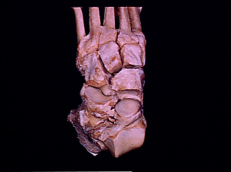
Joints of left ankle and foot
Joints of foot opened and viewed from above
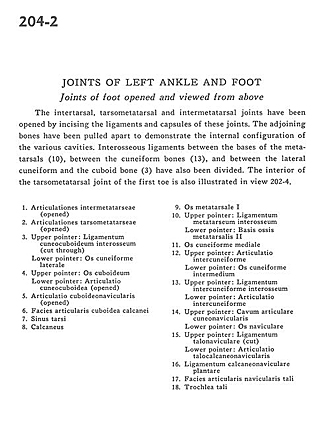
The intertarsal, tarsometatarsal and intermetatarsal joints have been opened by incising the ligaments and capsules of these joints. The adjoining bones have been pulled apart to demonstrate the internal configuration of the various cavities. Interosseous ligaments between the bases of the metatarsals (10), between the cuneiform bones (13), and between the lateral cuneiform and the cuboid bone (3) have also been divided. The interior of the tarsometatarsal joint of the first toe is also illustrated in view 202-4.
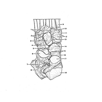
- Intermetatarsal articulations (opened)
- Tarsometatarsal articulations (opened)
- Upper pointer: Cuneocuboid interosseous ligament (cut through) Lower pointer: Lateral cuneiform bone
- Upper pointer: Cuboid bone Lower pointer: Cuneocuboid articulation (opened)
- Cuboideonavicular articulation (opened)
- Articular surface of calcaneus for cuboid
- Tarsal sinus
- Calcaneus
- Metatarsal bone
- Upper pointer: Metatarsal interosseous ligament Lower pointer: Base of second metatarsal bone
- Medial cuneiform bone
- Upper pointer: Intercuneiform articulation Lower pointer: Intermediate cuneiform bone
- Upper pointer: Intercuneiform interosseous ligament Lower pointer: Intercuneiform articulation
- Upper pointer: Cuneonavicular articular space Lower pointer: Navicular bone
- Upper pointer: Talonavicular ligament (cut) Lower pointer: Talocalcaneonavicular articulation
- Plantar calcaneonavicular ligament
- Articular surface of talus for navicular bone
- Trochlea of talus