
Bassett Collection of Stereoscopic Images of Human Anatomy
Joints of left ankle and foot
Interior of ankle joint and subtalar joint in relation to ligaments, posterior view
Image #203-5
KEYWORDS: Ankle, Bones joints cartilage, Foot and toes, Muscles and tendons.
Creative Commons
Stanford holds the copyright to the David L. Bassett anatomical images and has assigned Creative Commons license Attribution-Share Alike 4.0 International to all of the images.

For additional information regarding use and permissions, please contact the Medical History Center.

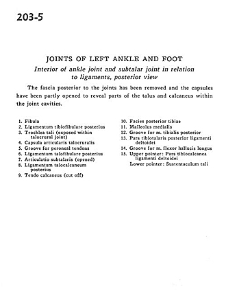
Joints of left ankle and foot
Interior of ankle joint and subtalar joint in relation to ligaments, posterior view
The fascia posterior to the joints has been removed and the capsules have been partly opened to reveal parts of the talus and calcaneus within the joint cavities.
- Fibula
- Posterior tibiofibular ligament
- Trochlea of talus (exposed within talocrural joint)
- Talocrural articular capsule
- Groove for peroneal tendons
- Posterior talofibular ligament
- Subtalar articulation (opened)
- Posterior talocalcaneal ligament
- Tendo calcaneus (cut off)
- Posterior surface of tibia
- Medial malleolus
- Groove for tibialis posterior muscle
- Posterior tibiotalar part of deltoid ligament
- Groove for flexor hallucis longus muscle
- Upper pointer: Tibiocalcaneal part of deltoid ligament Lower pointer: Sustentaculum tali