Bassett Collection of Stereoscopic Images of Human Anatomy
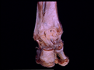
Joints of left ankle and foot
Ligaments of ankle and subtalar joints, posterior view
Image #203-4
KEYWORDS: Ankle, Bones joints cartilage, Foot and toes, Muscles and tendons.
Creative Commons
Stanford holds the copyright to the David L. Bassett anatomical images and has assigned Creative Commons license Attribution-Share Alike 4.0 International to all of the images.

For additional information regarding use and permissions, please contact the Medical History Center.


Joints of left ankle and foot
Ligaments of ankle and subtalar joints, posterior view
The capsules of the ankle and subtalar (talocalcaneal)joints remain intact. In addition, a layer of fascia (5) has been retained posteriorly. This layer continued laterally into the superior peroneal retinaculum and blended medially into the flexor retinaculum.
- Fibula
- Posterior tibiofibular ligament
- Lateral malleolus
- Superior peroneal retinaculum (divided)
- Fascia posterior to ankle joint (this layer blended with superior peroneal retinaculum)
- Calcaneofibular ligament
- Subtalar articular capsule
- Inferior peroneal retinaculum
- Calcaneus
- Lateral tubercle process of calcaneus bone
- Medial malleolus
- Upper pointer: Groove for tendon of tibialis posterior muscle Lower pointer: Groove for tendon of flexor digitorum longus muscle
- Talocrural articular capsule
- Deltoid ligament
- Groove for tendon of flexor hallucis longus muscle
- Tibialis posterior muscle (cut off at insertion into tuberosity of navicular)
- Sustentaculum tali
- Metatarsal bone