Bassett Collection of Stereoscopic Images of Human Anatomy
Dissection of plantar aspect of left foot
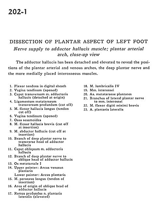
Nerve supply to adductor hallucis muscle; plantar arterial arch, close-up view
Image #202-1
KEYWORDS: Foot and toes, Muscles and tendons, Peripheral nervous system, Vasculature.
Creative Commons
Stanford holds the copyright to the David L. Bassett anatomical images and has assigned Creative Commons license Attribution-Share Alike 4.0 International to all of the images.

For additional information regarding use and permissions, please contact the Medical History Center.

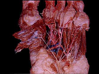
Dissection of plantar aspect of left foot
Nerve supply to adductor hallucis muscle; plantar arterial arch, close-up view
The adductor hallucis has been detached and elevated to reveal the positions of the plantar arterial and venous arches, the deep plantar nerve and the more medially placed interosseous muscles.
- Flexor tendons in digital sheath
- Tendinous sheath (opened)
- Transverse head of adductor hallucis muscle (detached at origin)
- Deep transverse metatarsal ligament (cut off)
- Flexor hallucis longus muscle (tendon cut off)
- Tendinous sheath (opened)
- Sesamoid bone
- Flexor hallucis brevis muscle (cut off at insertion)
- Abductor hallucis muscle (cut off at insertion)
- Branch of deep plantar nerve to transverse head of adductor hallucis
- Oblique head of adductor hallucis muscle
- Branch of deep plantar nerve to oblique head of adductor hallucis
- Metatarsal bone
- Upper pointer: Plantar venous arch Lower pointer: Plantar arch
- Peroneus longus muscle (tendon of insertion)
- Area of origin of oblique head of adductor hallucis
- Deep branch lateral plantar nerve (elevated)
- 4th lumbrical muscle
- Interosseous muscle
- Metatarsal plantar arteries
- Branches of lateral plantar nerve to interosseous muscle
- Flexor digiti minimi brevis muscle
- Lateral plantar artery