Bassett Collection of Stereoscopic Images of Human Anatomy
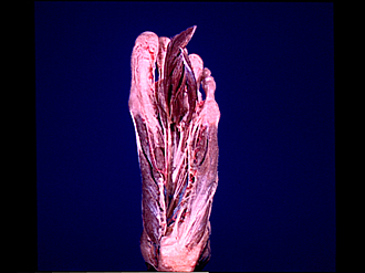
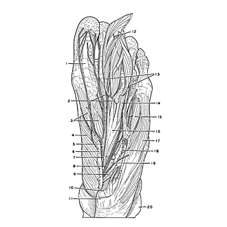
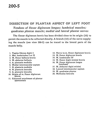
Dissection of plantar aspect of left foot
Tendons of flexor digitorum longus; lumbrical muscles; quadratus plantae muscle; medial and lateral plantar nerves
Image #200-5
KEYWORDS: Foot and toes, Muscles and tendons, Peripheral nervous system.
Creative Commons
Stanford holds the copyright to the David L. Bassett anatomical images and has assigned Creative Commons license Attribution-Share Alike 4.0 International to all of the images.

For additional information regarding use and permissions, please contact the Medical History Center.
Dissection of plantar aspect of left foot
Tendons of flexor digitorum longus; lumbrical muscles; quadratus plantae muscle; medial and lateral plantar nerves
The flexor digitorum brevis has been divided close to its origin (10) to permit the muscle to be reflected distally. A branch (12) of the nerve supplying the muscle (see view 200-2) can be traced to the lateral parts of the muscle belly.
- Fibrous sheath 1st digit
- 1st and 3rd lumbrical muscles
- Flexor hallucis brevis muscle
- Abductor hallucis muscle
- Medial plantar artery
- Medial intermuscular septum
- Medial plantar nerve
- Lateral plantar nerve
- Lateral plantar artery
- Origin of flexor digitorum brevis muscle
- Calcaneal attachment of plantar aponeurosis
- Nerve to flexor digitorum brevis muscle
- Flexor digitorum brevis muscle
- 4th lumbrical muscle
- Flexor digiti minimi brevis muscle
- Flexor digitorum longus muscle (tendons)
- Abductor digiti minimi muscle
- Lateral intermuscular septum
- Quadratus plantae muscle
- Lateral malleolus