
Bassett Collection of Stereoscopic Images of Human Anatomy
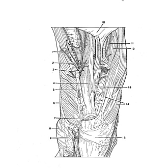
Dissection of plantar aspect of left foot
Abductor hallucis, flexor digitorum brevis and abductor digiti minimi muscles; nerve supply to flexor digitorum brevis
Image #200-2
KEYWORDS: Foot and toes, Muscles and tendons, Peripheral nervous system.
Creative Commons
Stanford holds the copyright to the David L. Bassett anatomical images and has assigned Creative Commons license Attribution-Share Alike 4.0 International to all of the images.

For additional information regarding use and permissions, please contact the Medical History Center.

Dissection of plantar aspect of left foot
Abductor hallucis, flexor digitorum brevis and abductor digiti minimi muscles; nerve supply to flexor digitorum brevis
The plantar aponeurosis (10) has been reflected toward the toes. Its attachments to intermuscular septa are visible in several places. The nerve to the flexor digitorum brevis (3) enters the plantar surface of the muscle and is visible in the dissection.
- Flexor hallucis brevis muscle
- Branch of medial plantar artery
- Nerve to flexor digitorum brevis muscle
- Flexor digitorum brevis muscle
- Medial intermuscular septum
- Abductor hallucis muscle
- Plantar aponeurosis (cut at attachment to calcaneus)
- Medial malleolus
- Flexor retinaculum
- Plantar aponeurosis (reflected)
- Common plantar digital nerve
- Flexor digiti minimi brevis muscle
- Lateral intermuscular septum
- Abductor digiti minimi muscle
- Tuberosity of calcaneus