Bassett Collection of Stereoscopic Images of Human Anatomy
Exploration of those parts of the brain supplied by the posterior cerebral artery
Tapetum and tail of caudate nucleus
Image #20-4
KEYWORDS: Brain, Telencephalon.
Creative Commons
Stanford holds the copyright to the David L. Bassett anatomical images and has assigned Creative Commons license Attribution-Share Alike 4.0 International to all of the images.

For additional information regarding use and permissions, please contact the Medical History Center.
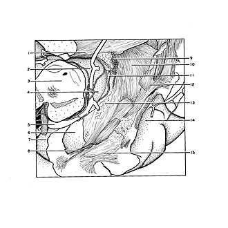

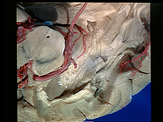
Exploration of those parts of the brain supplied by the posterior cerebral artery
Tapetum and tail of caudate nucleus
The ependymal lining has been removed to expose the tapetum (10), a broad band of fibers sweeping downward from the corpus callosum over the inferior horn of the ventricle into the temporal lobe. As the tail of the caudate nucleus (13) passes down from the floor of the central part of the ventricle into the roof of the inferior horn it parallels the anterior edge of the tapetum (left border as seen in the view). Only a fragmentary part of the tail of the caudate nucleus could be satisfactorily demonstrated in this specimen.
- Corpus callosum (splenium)
- Pulvinar
- Mesencephalon
- Lateral geniculate body
- Posterior cerebral artery
- Amygdaloid nucleus (ventricular surface)
- Uncus
- Fibers passing beneath lateral ventricle into uncus
- Choroid plexus
- Tapetum
- Stria terminalis
- Medullary substance of occipital lobe
- Caudate nucleus (tail)
- Inferior temporal sulcus
- Medullary substance of temporal lobe