Bassett Collection of Stereoscopic Images of Human Anatomy
Exploration of the meninges and brain in situ
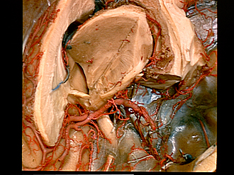
Optic nerve, optic chiasm, optic tract and lateral geniculate body viewed from above and in front
Image #2-7
KEYWORDS: Brain, Diencephalon, Peripheral nervous system, Telencephalon, Vasculature.
Creative Commons
Stanford holds the copyright to the David L. Bassett anatomical images and has assigned Creative Commons license Attribution-Share Alike 4.0 International to all of the images.
For additional information regarding use and permissions, please contact the Medical History Center.
Exploration of the meninges and brain in situ
Optic nerve, optic chiasm, optic tract and lateral geniculate body viewed from above and in front
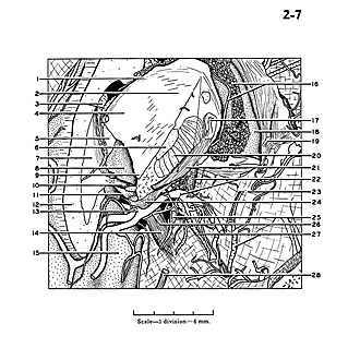
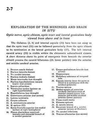
The thalamus (2, 4) and internal capsule (20) have been cut away so that the optic tract (21) can be followed posteriorly from the optic chiasm to its termination at the lateral geniculate body (17). The left internal carotid artery (25) is visible within the chiasmatic subarachnoid cistern. A short distance above its point of emergence from beneath the anterior clinoid process the carotid bifurcates (22, lower pointer) into the anterior and middle cerebral arteries.
- Striatum zone of thalamus
- Lateral nucleus of thalamus
- Internal cerebral vein
- Medial nucleus of thalamus
- Massa intermedia (cut through)
- Hypothalamus (cut across)
- Septum pellucidum
- Third ventricle (pointer on right hypothalamus)
- Hypothalamus (cut through)
- Anterior commissure (cut across)
- Lamina terminalis
- Corpus callosum
- Anterior communicating artery
- Optic nerve (11)
- Superior frontal gyrus (on medial surface of right hemisphere)
- Choroid plexus lateral ventricle and choroidal branch of posterior cerebral artery
- Lateral geniculate body (cut across)
- Hippocampus
- Medullary substance of temporal lobe
- Internal capsule (near the point at which it is continuous with the cerebral peduncle)
- Optic tract
- Medial margin of tentorium cerebelli and bifurcation of internal carotid artery into anterior and middle cerebral arteries (lower pointer)
- Middle cranial fossa
- Optic chiasm
- Internal carotid artery
- Recurrent branch of anterior cerebral artery (artery of Heubner)
- Inferior cerebral vein
- Olfactory tract (cut across)


