Bassett Collection of Stereoscopic Images of Human Anatomy
Exploration of the meninges and brain in situ
Pericallosal arteries (both arising from right anterior cerebral artery)
Image #2-2
KEYWORDS: Brain, Meninges, Telencephalon, Vasculature.
Creative Commons
Stanford holds the copyright to the David L. Bassett anatomical images and has assigned Creative Commons license Attribution-Share Alike 4.0 International to all of the images.

For additional information regarding use and permissions, please contact the Medical History Center.

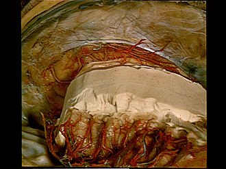
Exploration of the meninges and brain in situ
Pericallosal arteries (both arising from right anterior cerebral artery)
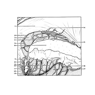
The left anterior cerebral artery has been removed. The pericallosal arteries, normally terminal branches of the anterior cerebral arteries of their respective sides, both arise from the right side in this specimen.
- Falx cerebri
- Cingulate sulcus
- Cingulate gyrus
- Corpus callosum (trunk)
- Callosomarginal branch of right anterior cerebral artery
- Corona radiata and medullaiy substance of hemisphere (cut across)
- Sulcus circularis at margin of insula
- Arachnoid lifted up to display the digital impressions of the orbital roof (dura intact)
- Position of crista galli to which falx cerebri attaches
- Short insular gyrus
- Branches of middle cerebral artery
- Digital impressions in floor of anterior cranial fossa
- Lateral fissure (Sylvian)
- Right and left pericallosal arteries both arising from right anterior cerebral artery
- Corpus callosum (splenium)
- Posterior parietal branch of middle cerebral artery
- Transverse temporal gyrus