Bassett Collection of Stereoscopic Images of Human Anatomy
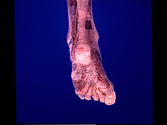
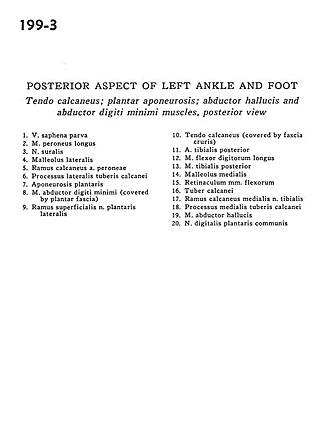
Posterior aspect of left ankle and foot
Tendo calcaneus; plantar aponeurosis; abductor hallucis and abductor digiti minimi muscles, posterior view
Image #199-3
KEYWORDS: Ankle, Foot and toes, Muscles and tendons.
Creative Commons
Stanford holds the copyright to the David L. Bassett anatomical images and has assigned Creative Commons license Attribution-Share Alike 4.0 International to all of the images.

For additional information regarding use and permissions, please contact the Medical History Center.
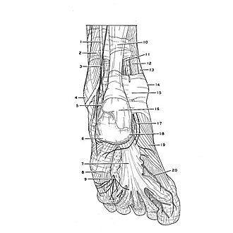
Posterior aspect of left ankle and foot
Tendo calcaneus; plantar aponeurosis; abductor hallucis and abductor digiti minimi muscles, posterior view
- Lesser saphenous vein
- Peroneus longus muscle
- Sural nerve
- Lateral malleolus
- Calcaneal branch of peroneal artery
- Lateral tubercle process of calcaneus bone
- Plantar aponeurosis
- Abductor digiti minimi muscle (covered by plantar fascia)
- Superficial branch of lateral plantar nerve
- Tendo calcaneus (covered by crural fascia)
- Posterior tibial artery
- Flexor digitorum longus muscle
- Tibialis posterior muscle
- Medial malleolus
- Flexor retinaculum
- Tuberosity of calcaneus
- Calcaneal branch of medial tibial nerve
- Medial process of tuberosity of calcaneus
- Abductor hallucis muscle
- Common plantar digital nerve