Bassett Collection of Stereoscopic Images of Human Anatomy
Dissection of medial aspect of left foot and ankle
Long flexor tendons within flexor retinaculum, medial view of ankle
Image #199-2
KEYWORDS: Ankle, Foot and toes, Muscles and tendons.
Creative Commons
Stanford holds the copyright to the David L. Bassett anatomical images and has assigned Creative Commons license Attribution-Share Alike 4.0 International to all of the images.

For additional information regarding use and permissions, please contact the Medical History Center.
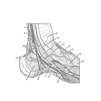
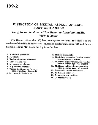
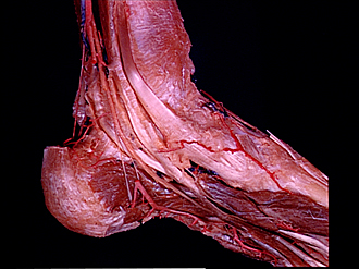
Dissection of medial aspect of left foot and ankle
Long flexor tendons within flexor retinaculum, medial view of ankle
The flexor retinaculum (3) has been opened to reveal the course of the tendons of the tibialis posterior (10), flexor digitorum longus (11) and flexor hallucis longus (12) from the leg into the foot.
- Posterior tibial artery
- Tibial nerve
- Flexor retinaculum
- Tendo calcaneus
- Quadratus plantae muscle
- Lateral plantar artery
- Tendinous sheath of flexor digitorum longus (opened)
- Flexor hallucis brevis muscle
- Medial malleolus
- Tibialis posterior muscle (tendon within opened synovial sheath)
- Flexor digitorum longus muscle (tendon within opened synovial sheath)
- Flexor hallucis longus muscle (tendon within opened synovial sheath)
- Tuberosity of navicular bone
- Tibialis anterior muscle
- Medial cuneiform bone
- metatarsal bone