Bassett Collection of Stereoscopic Images of Human Anatomy
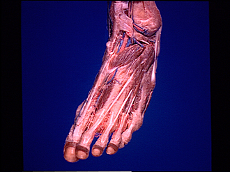
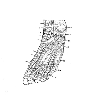
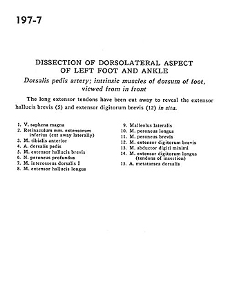
Dissection of dorsolateral aspect of left foot and ankle
Dorsalis pedis artery; intrinsic muscles of dorsum of foot, viewed from in front
Image #197-7
KEYWORDS: Ankle, Foot and toes, Muscles and tendons, Vasculature.
Creative Commons
Stanford holds the copyright to the David L. Bassett anatomical images and has assigned Creative Commons license Attribution-Share Alike 4.0 International to all of the images.

For additional information regarding use and permissions, please contact the Medical History Center.
Dissection of dorsolateral aspect of left foot and ankle
Dorsalis pedis artery; intrinsic muscles of dorsum of foot, viewed from in front
The long extensor tendons have been cut away to reveal the extensor hallucis brevis (5) and extensor digitorum brevis (12) in situ.
- Greater saphenous vein
- Inferior extensor retinaculum (cut away laterally)
- Tibialis anterior muscle
- Dorsalis pedis artery
- Extensor hallucis brevis muscle
- Deep peroneal nerve
- 1st dorsal interosseus muscle
- Extensor hallucis longus muscle
- Lateral malleolus
- Peroneus longus muscle
- Peroneus brevis muscle
- Extensor digitorum brevis muscle
- Abductor digiti minimi muscle
- Extensor digitorum longus muscle (tendons of insertion)
- Dorsal metatarsal artery