
Bassett Collection of Stereoscopic Images of Human Anatomy
Dissection of dorsolateral aspect of left foot and ankle
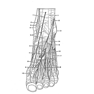
Superficial nerves and vessels of dorsum of foot in relation to muscles and tendons, viewed from in front
Image #197-1
KEYWORDS: Ankle, Foot and toes, Muscles and tendons, Peripheral nervous system, Vasculature.
Creative Commons
Stanford holds the copyright to the David L. Bassett anatomical images and has assigned Creative Commons license Attribution-Share Alike 4.0 International to all of the images.

For additional information regarding use and permissions, please contact the Medical History Center.

Dissection of dorsolateral aspect of left foot and ankle
Superficial nerves and vessels of dorsum of foot in relation to muscles and tendons, viewed from in front
The superficial nerves have been preserved along with the major superficial venous pathways. The dorsal fascia of the foot has been partially removed.
- Dorsal medial cutaneous nerve
- Greater saphenous vein
- Saphenous nerve
- Medial malleolus
- Inferior extensor retinaculum
- Extensor hallucis brevis muscle
- Dorsalis pedis artery
- Venous arch of dorsalis pedis
- Deep peroneal nerve
- Extensor hallucis longus muscle (tendon)
- Superior extensor retinaculum
- Dorsal intermediate cutaneous nerve
- Lateral malleolus
- Tendinous sheath of extensor digitorum longus muscle
- Extensor digitorum brevis muscle
- Branch of deep peroneal nerve to lateral side of second toe
- Fascia of dorsalis pedis
- Extensor digitorum brevis muscle (tendons)