Bassett Collection of Stereoscopic Images of Human Anatomy
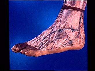
Dissection of dorsolateral aspect of left foot and ankle
Superficial nerves and blood vessels of dorsum of foot
Image #196-6
KEYWORDS: Ankle, Foot and toes, Peripheral nervous system, Vasculature.
Creative Commons
Stanford holds the copyright to the David L. Bassett anatomical images and has assigned Creative Commons license Attribution-Share Alike 4.0 International to all of the images.

For additional information regarding use and permissions, please contact the Medical History Center.

Dissection of dorsolateral aspect of left foot and ankle
Superficial nerves and blood vessels of dorsum of foot
A margin of intact skin and tela subcutanea has been retained surrounding the area of the foot and ankle which has been dissected. The cutaneous nerves and superficial veins have been exposed by dissection of the tela subcutanea. The deep fascia has not been disturbed.
- Dorsal intermediate cutaneous nerve
- Dorsal medial cutaneous nerve
- Fascia of dorsalis pedis (overlying extensor digitorum brevis muscle)
- Tendons of extensor digitorum longus muscle (covered by fascia)
- Venous arch of dorsalis pedis
- Metatarsal veins of dorsalis pedis
- Lesser saphenous vein
- Lateral malleolus
- Sural nerve (continuing on foot as dorsal lateral cutaneous nerve)
- Tela subcutanea
- Venous network
- Dorsal digital nerves