Bassett Collection of Stereoscopic Images of Human Anatomy
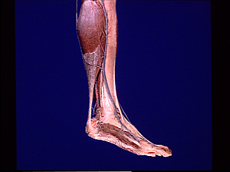
Dissection of medial aspect of left leg
Large saphenous vein; muscles of leg and foot, medial view
Image #194-2
KEYWORDS: Foot and toes, Leg, Muscles and tendons, Peripheral nervous system, Vasculature.
Creative Commons
Stanford holds the copyright to the David L. Bassett anatomical images and has assigned Creative Commons license Attribution-Share Alike 4.0 International to all of the images.

For additional information regarding use and permissions, please contact the Medical History Center.


Dissection of medial aspect of left leg
Large saphenous vein; muscles of leg and foot, medial view
The deep fascia of the leg and foot has been removed. However, the superficial nerves (1,7,18) and the large saphenous vein (11) have beem retained to illustrate their relations to the deeper structures.
- Saphenous nerve (traceable to great toe)
- Gastrocnemius muscle
- Posterior tibial artery
- Tendo calcaneus (Achilis)
- Tibial nerve
- Flexor retinaculum
- Medial calcaneal branches of tibial nerve
- Calcaneus
- Abductor hallucis muscle
- Plantar aponeurosis
- Greater saphenous vein
- Soleus muscle
- Medial surface of tibia
- Flexor digitorum longus muscle
- Tibialis posterior muscle
- Tibialis anterior muscle (tendon, covered by superior retinaculum)
- Medial malleolus
- Dorsal medial cutaneous nerve
- Extensor hallucis longus muscle (tendon)